
Introduction to Peripheral Nervous System (PNS)
Peripheral Nervous System is comprised of two nervous systems : Somatic nervous system (SNS) and Autonomic Nervous System (ANS). Even though Central Nervous System (CNS) is considered as master of perception and behaviour, Peripheral Nervous System is the one who co-ordinates and maintains the fundamental bodily functions. SNS contains sensory and motor nerves that extends all over the body that connects the CNS (brain and spinal cord) to the target organs. While ANS handles the vital functions like circulation, breathing, hormone secretion; that too without consciousness.
The peripheral nervous system (PNS) comprises the nerves and ganglia that lie outside the central nervous system (CNS), which includes the brain and spinal cord. The term “peripheral” originates from the Latin word peripheria (meaning “outer edge” or “circumference”), reflecting its role in extending to the outer regions of the body and connecting the CNS to the limbs, organs, and tissues.
Somatic Nervous System (SNS)
SNS mainly comprises of nerves extending all over the body, originating primarily from CNS (Smith, 2017). There are two main types of nerves based on their origin.
Cranial nerves :
There 12 pairs of nerves that originate from brain that controls the head and neck region as well as various internal organs. They are of both types – purely sensory as well as sensory plus motor in nature. They are numbered in sequence from front to back, and each nerve has the special function. They are ipsilateral means set of nerve present on left side handles functioning of left side and the nerves on right side work for right side of the body.
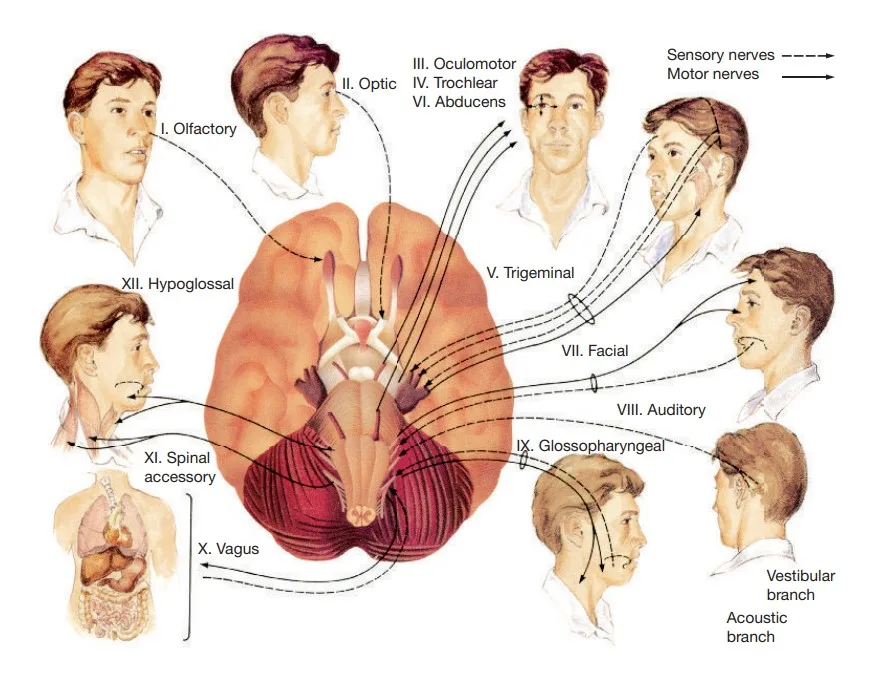
Cranial nerves. J. Pinel & S. Barnes (2018). Biopsychology. 10th ed.
The longest cranial nerve is Vagus nerve which extends till the guts.
Function of cranial nerves :
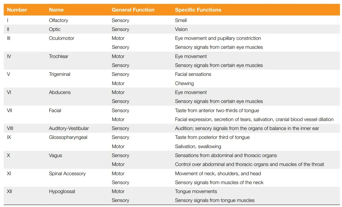
Functions of cranial nerves. J. Pinel & S. Barnes (2018). Biopsychology. 10th ed.
2. Spinal nerves :
The 31 pairs of nerves that originate from the spinal cord are known as spinal nerves. They are divided into 4 types based on the corresponding division of spinal cord from where they are originated. They are cervical nerves, thoracic nerves, lumbar nerves and sacral nerves. For the effective functioning, the body is divided into the segments called as “dermatomes” (meaning skin cut) and regulated by specific set of spinal nerves.
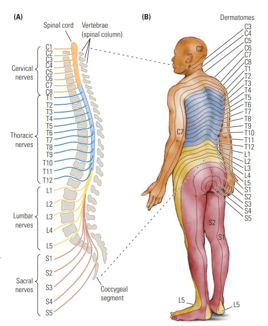
Spinal cord and spinal nerve structure. J. Pinel & S. Barnes (2018). Biopsychology. 10th ed.
Spinal nerves have mixed nerve structure i.e they’re sensory as well as motor in nature. A dermatome has a sensory nerve to send information from the skin, joints, and muscles to the spinal cord, as well as a motor nerve to control the muscle movements in that particular body segment. As cranial nerves receive information from sensory receptors in the eyes, ears, facial skin, and so forth, the spinal nerves receive information from sensory receptors in the rest of the body.
Learn the internal structure of spinal cord
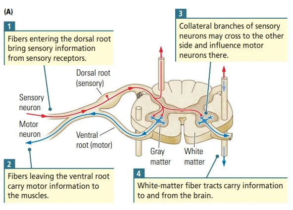
Cross section of Spinal cord. Kolb and Whishaw (2009). Fundamentals of Human Neuropsychology
- Sensory function (Afferent Pathways):
- Spinal nerves carry sensory information from the body (e.g., skin, muscles, joints, and internal organs) to the CNS.
- They transmit signals such as pain, temperature, touch, and proprioception (awareness of body position).
- Motor Function (Efferent Pathways):
- Spinal nerves convey motor commands from the CNS to skeletal muscles, enabling voluntary movements.
- They also transmit autonomic motor signals to smooth muscles, glands, and other structures.
- Reflex Action:
- Spinal nerves are essential in reflex arcs, which are automatic and rapid responses to stimuli. For example, the patellar reflex involves a spinal nerve mediating a motor response to a sensory input without involving the brain.
- Regional Specialization:
- Cervical spinal nerves: Control the neck, diaphragm (via the phrenic nerve), arms, and shoulders.
- Thoracic spinal nerves: Innervate the chest and abdominal muscles.
- Lumbar spinal nerves: Control the lower back, hips, and parts of the legs.
- Sacral spinal nerves: Manage bowel, bladder, and reproductive organs, along with parts of the legs.
- Coccygeal nerve: Plays a minor role in sensory functions of the tailbone area.
Autonomic Nervous System (ANS)
The autonomic nervous system (ANS) is a division of the peripheral nervous system (PNS) that regulates involuntary physiological processes in the body. It operates largely without conscious control and is responsible for maintaining homeostasis by controlling functions such as heart rate, blood pressure, digestion, respiration, and body temperature.
Divisions of the Autonomic Nervous System
The ANS has two primary branches that work together to maintain balance in the body’s internal environment:
Sympathetic Nervous System (SNS):
- Known as the “fight-or-flight” system.
- Prepares the body for stressful or emergency situations by increasing heart rate, dilating airways, and inhibiting non-essential functions like digestion.
- Example: When you face danger, the SNS triggers a rapid response to prepare you for action.
Parasympathetic Nervous System (PNS):
- Known as the “rest-and-digest” system.
- Promotes relaxation and conservation of energy by slowing heart rate, stimulating digestion, and enhancing recovery processes.
- Example: After a meal, the PNS activates to support digestion and nutrient absorption.
Function of ANS
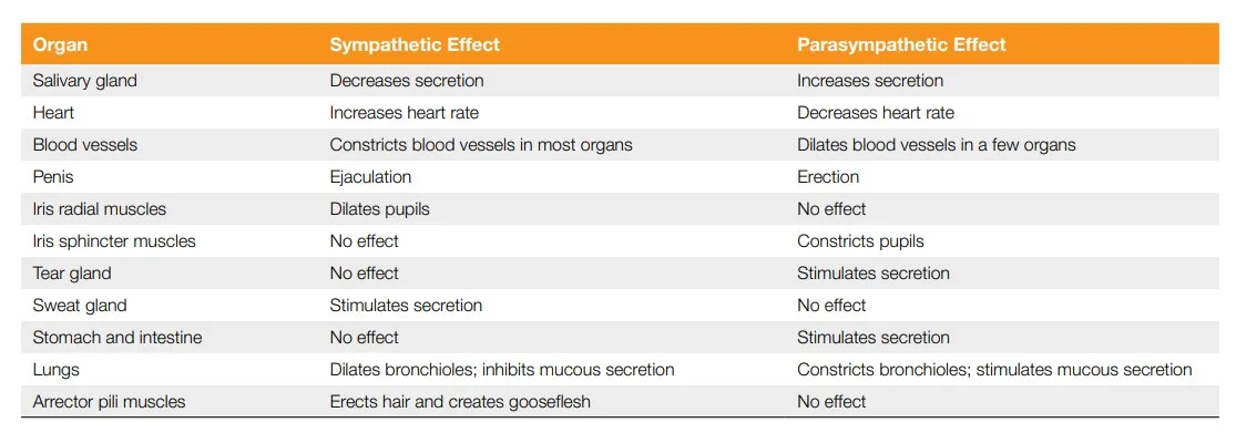
Functions of ANS. J. Pinel & S. Barnes (2018). Biopsychology. 10th ed.
Ganglia
Ganglia are critical components of the autonomic nervous system (ANS). Ganglia are defined as Spherical or fusiform cluster of sensory nerve cell bodies in the PNS or of small motor nerve cell bodies in the ANS which has connective tissue investments like those of adjoining nerves (Angevine, 2002).
Ganglia serve as relay hubs for autonomic signals from the spinal cord or cranial nerves. There are two types of ganglia :
- Sympathetic ganglia : Sympathetic ganglia are located near the spinal cord, forming sympathetic chains on either side of the spinal cord. These ganglia act as “mini-brains” for regulating specific internal organs. The activation of ANS starts in the thoracic and lumbar regions of the spinal cord. It is mediated to respective internal organs via sympathetic chain ganglia which later clusters into 3 sympathetic prevertebral ganglia.
2. Parasympathetic ganglia : Parasympathetic ganglia are located near target organ and connected to the sacral region of the spinal cord & three cranial nerves:
- Vagus Nerve (Cranial Nerve X): Calms most internal organs (e.g., slows heart rate, stimulates digestion).
- Facial Nerve (Cranial Nerve VII): Controls salivation.
- Oculomotor Nerve (Cranial Nerve III): Controls pupil constriction and lens adjustment.

Autonomic Nervous system
Ganglia allow the ANS to function semi-independently of the CNS for certain reflexive and local processes.
1. Preganglionic Neuron
- Origin: The cell body of a preganglionic neuron is located in the CNS (either in the brainstem or spinal cord).
- Pathway: Its axon extends outward to synapse in a ganglion (a cluster of neuronal cell bodies in the PNS).
- Neurotransmitter: Preganglionic neurons release acetylcholine (ACh) to stimulate the postganglionic neuron.
2. Postganglionic Neuron
- Origin: The cell body of a postganglionic neuron resides in the ganglion.
- Pathway: Its axon extends from the ganglion to the target organ (e.g., heart, lungs, digestive tract).
- Neurotransmitter:
- Sympathetic division: Typically releases norepinephrine (NE), except in sweat glands where it releases acetylcholine.
- Parasympathetic division: Releases acetylcholine (ACh).
Enteric Nervous System (ENS)
The Enteric Nervous System (ENS) is a fascinating and complex component of the nervous system, often referred to as the “second brain” due to its autonomy and intricate functions in managing gut activity. Although part of the autonomic nervous system (ANS), the ENS largely functions independently of the central nervous system (CNS). It manages complex digestive processes like motility, secretion, blood flow, and nutrient absorption.
Some scientists propose that the ENS represents an evolutionary precursor to the CNS, emphasizing its advanced neuronal architecture and functional independence.
🟢 Structure and Neuronal Network:
- Comprises 200–500 million neurons, similar in number to the spinal cord.
- ENS neurons are organized into two layers of ganglia in the gut lining:
- Myenteric Plexus: Controls gut motility.
- Submucosal Plexus: Regulates secretion and blood flow.
- Neurons and glial cells form ganglia interconnected by nerve fibers.
🟢 Chemical and Hormonal Sensitivity: The ENS responds to a variety of hormones and chemicals, ensuring efficient digestion and waste elimination.
🟢 Connection to the CNS:
- Linked to the brain via the ANS, particularly the vagus nerve, facilitating bidirectional communication.
- Stress and emotions can alter ENS function, contributing to symptoms like nausea or diarrhea.
🟢 Role of the Gut Microbiota:
- Microbiome and Neurochemicals: The gut hosts approximately 3.9 × 10¹³ microbiota, outnumbering human cells. These microorganisms influence digestion, nutrient absorption, and neurochemical production, affecting both physiological and psychological processes.
- Psychobiotics: Emerging compounds that leverage live microorganisms to address behavioral and psychological disorders by influencing the ENS and CNS.
🟢 Impact on Behavior and Mental State:
- ENS and Mental Health: The ENS communicates directly with the brain, influencing mental states. Stress and anxiety impact gut function, and gut signals can reciprocally affect mood and behavior.
- Behavioral Disorders: Evidence suggests ENS involvement in behavioral conditions, with treatments focusing on modulating the microbiota.
Conclusion
The peripheral nervous system (PNS) is a vital network that bridges the central nervous system (CNS) with the rest of the body, ensuring efficient communication and control over voluntary and involuntary functions. It encompasses three major subdivisions: the somatic nervous system (SNS), autonomic nervous system (ANS), and enteric nervous system (ENS), each playing distinct yet interconnected roles.
Together, these systems exemplify the complexity of the PNS, balancing conscious control, automatic regulation, and independent digestive functions. This seamless integration highlights the PNS’s indispensable role in maintaining physiological harmony and supporting adaptive responses to both internal and external challenges.
Learn more about Neuropsychology
References
Angevine, J. B. (2002). Nervous System, Organization of. In Elsevier eBooks (pp. 313–371). https://doi.org/10.1016/b0-12-227210-2/00235-1
Kalat, J. W. (2019). Biological psychology. Cengage.
Kolb, B., & Whishaw, I. Q. (2019). An introduction to brain and behavior.
Pinel, J. (2023). Biopsychology 10th Edition. Pearson.
Smith, S. A. (2017). Acquired peripheral neuropathies. In Elsevier eBooks (pp. 1081–1085). https://doi.org/10.1016/b978-0-323-37101-8.00142-9
Niwlikar, B. A. (2025, January 23). 2 Types of Peripheral Nervous System : SNS & ANS. Careershodh. https://www.careershodh.com/2-types-of-peripheral-nervous-system-sns-ans/