
Introduction to Structure of Neurons
Neuron is considered as the basic cellular unit of the nervous system. As a psychologist, we are interested in study of nervous system, structure of neurons and their functioning as it helps to bridge the gap between behavior and biology. It enables to understand the mechanism behind mental processes and behaviors initiated by it more holistically. It allows to better comprehend the biological basis of mental processes and disorders, thereby improving interventions and treatments.
To understand the Nervous system more deeply, let’s begin with the smallest part of it i.e NEURON.
Definition of Neuron
- Neurons are the information-conducting units of the nervous system. (Kolb B. & Whishaw I., 2008)
- National Institute of Cancer (NIH) defines neuron as, “A type of cell that receives and sends messages from the body to the brain and back to the body. The messages are sent by a weak electrical current. Also called nerve cell.”
The neuron is also known as Nerve cell, the term coined by German physician Heinrich Wilhelm von Waldeyer-Hartz (1836–1921).
Structure of Neuron
The neuron has 3 main parts : Body (soma), Dendrites and Axon.
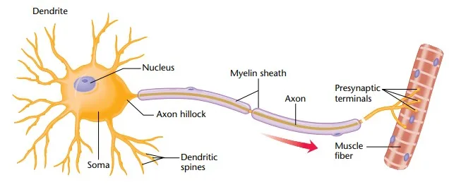
Structure of neuron
1st structure of Neuron : Body (Soma)
The cell body, also known as the soma (from the Greek word for “body”), is the central part of the neuron that contains essential organelles for maintaining its functions. It houses the nucleus, ribosomes, mitochondria, and other structures found in most cells, where much of the metabolic work of the nerve cell takes place.
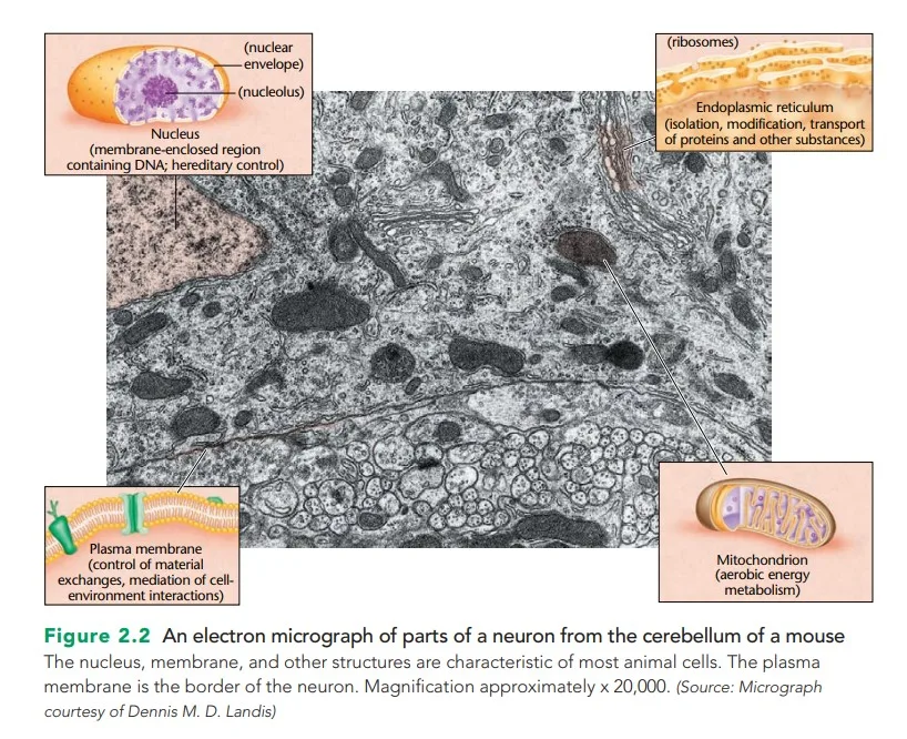
Soma of Neuron
Structural Features
- Nucleus:
- Often referred to as the “executive office” of the neuron, the nucleus houses the blueprints of life—genes and chromosomes.
- These genetic instructions dictate the synthesis of proteins, crucial for cellular functions and communication.
- Copies of genetic instructions are sent to the endoplasmic reticulum (ER) for protein assembly.
- Endoplasmic Reticulum (ER):
- The ER is an extension of the nuclear membrane where proteins are synthesized according to genetic codes.
- Proteins are then packaged in membranes by the Golgi apparatus and transported to their destinations via a system of tubules—similar to conveyor belts in a factory.
- Ribosomes :
- Ribosomes are the sites at which the cell synthesizes new protein molecules. Proteins provide building materials for the cell and facilitate various chemical reactions. Some ribosomes float freely within the cell, while some ribosomes are attached to ER.
- Mitochondria:
- These are the “powerhouse” of the neuron, providing the energy required for its metabolic needs.
- Energy is critical for the neuron’s functions, such as transmitting electrical signals and maintaining ion gradients.
- Lysosomes:
- These saclike vesicles transport supplies within the cell and store or dispose of waste materials.
- Interestingly, older nerves contain more lysosomes, reflecting an accumulation of waste over time.
- Cytoskeletal Framework:
- Microfilaments and microtubules form the structural framework of the nerve cell.
- These structures not only provide stability but also assist in cellular movements and intracellular transport of substances
Size of Body of neuron :
- Mammalian neurons range from 0.005 mm to 0.1 mm in diameter.
- Some invertebrates can have cell bodies as large as 1 mm.
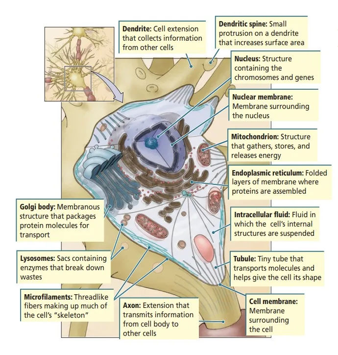
Internal structure of neuron’s body
2nd structure of neuron : Dendrites
Dendrites are branching, tree-like extensions of a neuron that serve as the primary sites for receiving signals from other nerve cells. The term “dendrite” is derived from the Greek word for “tree,” reflecting their branched appearance. These structures play a vital role in neuronal communication by gathering and processing inputs. A single neuron can have 1 to 20 dendrites, each branching extensively.
Structural Features:
- Branching Fibers:
- Dendrites extend outward from the cell body and taper as they branch, resembling the branches of a tree.
- Their extensive branching increases the neuron’s surface area, allowing it to receive more information.
- Synaptic Receptors:
- The dendritic surface is lined with synaptic receptors, specialized sites where chemical signals from other neurons are received.
- These receptors translate incoming signals into electrical or biochemical responses within the neuron.
- Dendritic Spines:
- Many dendrites are adorned with dendritic spines, which are small protrusions that further increase the surface area for synaptic connections.
- Each nerve cell can have thousands of spines, providing numerous points of communication. The presence of these spines indicates the neuron’s capacity to process and store large amounts of information.
- The density and shape of these spines can change over time, affecting how the nerve cell processes information.
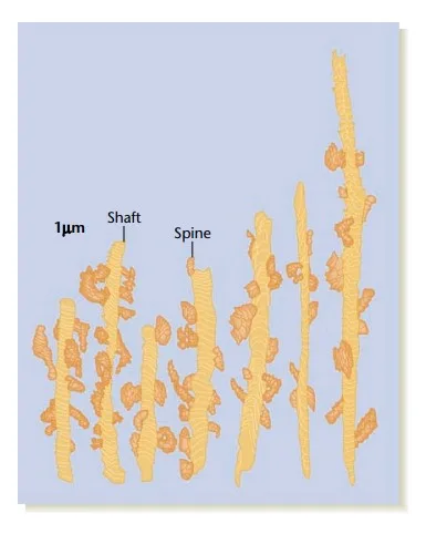
Dendritic spines. (Source: From K. M. Harris and J. K. Stevens, Society for Neuroscience, “Dendritic spines of CA1 pyramidal cells in the rat hippocampus: Serial electron microscopy with reference to their biophysical characteristics.” Journal of Neuroscience, 9, 1989,
2982–2997.)
Variability in Shape:
- The structure of dendrites varies widely across different types of neurons.
- Even within a single nerve cell, the shape of the dendrites can change over time in response to activity and environmental factors (Häusser, Spruston, & Stuart, 2000).
Functional Significance:
- Information Reception: Dendrites collect signals from other neurons through synapses. The larger the dendritic surface area, the greater the amount of information the nerve cell can receive and process.
- Integration of Inputs: The branching structure and shape of dendrites influence how a neuron integrates multiple types of input. This integration determines whether the neuron will generate an action potential to transmit information further.
- Plasticity: Dendrites are highly dynamic. The growth, retraction, or modification of dendritic spines underlies learning and memory, as these changes enhance or diminish synaptic connections.
3rd structure of neuron : Axon
The axon is a long, slender projection of a neuron that plays a crucial role in transmitting signals to other neurons, muscles, or glands. Its structure and function are uniquely suited for sending information, distinguishing it as the neuron’s information sender.
Structural Features:
Constant Diameter and Length: The axon maintains a constant diameter along its length and is often longer than the dendrites, with some axons in the human body extending over a meter, such as those running from the spinal cord to the feet.
- Axon Hillock: The axon originates from a region of the cell body called the axon hillock (“little hill”), where electrical signals are typically initiated.
- Branches and Terminals:
- The axon may have branches, known as axon collaterals, that extend at right angles.
- Toward the end, the axon divides into teleodendria (“end branches”), each ending in a terminal button or end foot, which plays a role in communication with the target cell.
- Myelin Sheath and Nodes of Ranvier:
- In vertebrates, many axons are insulated with a myelin sheath, a fatty layer that increases the speed of signal transmission.
- The sheath is interrupted by gaps called nodes of Ranvier, which facilitate rapid conduction of electrical impulses through a process called saltatory conduction.
- Invertebrate axons lack myelin sheaths but can transmit signals effectively due to their structural adaptations.
Functional Significance:
- Signal Transmission: The axon is responsible for transmitting electrical impulses, known as action potentials, away from the cell body toward the axon terminals.
- Synaptic Communication: At the terminal buttons, the axon releases neurotransmitters into the synaptic cleft—the small gap between the axon terminal and the dendrite of the next neuron. This process allows the nerve cell to communicate with its target cell.
- Directional Flow: Neurons have only one axon, limiting them to a single output channel for communication. This is in contrast to the multiple dendrites that gather input from various sources.
Types of Axons
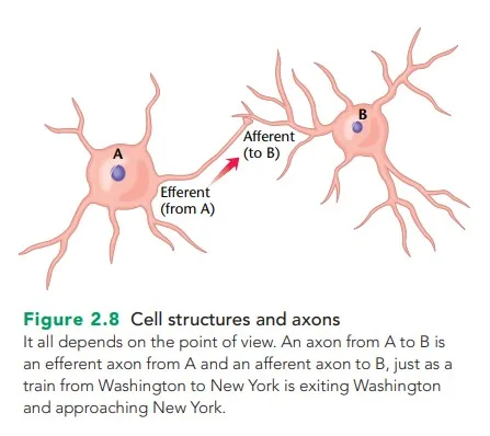
- Afferent and Efferent Axons:
- An afferent axon brings information into a structure (e.g., sensory neurons bringing signals to the nervous system).
- An efferent axon carries information away from a structure (e.g., motor neurons sending signals to muscles).
- A neuron may be efferent relative to one structure and afferent relative to another. For instance, an axon from the thalamus is efferent to the thalamus but afferent to the cerebral cortex.
- Interneurons (Intrinsic Neurons):
- These neurons have both dendrites and axons confined within a single structure, such as an interneuron in the thalamus that communicates only with other thalamic neurons.
Flow of Information Through the Neuron
Step 1: Reception at Dendrites
- Dendrites, with their numerous branches and dendritic spines, act as the neuron’s primary information-receiving structures.
- They are lined with synaptic receptors that collect incoming signals from other neurons.
- These signals are typically in the form of chemical messages released by the axons of neighbouring nerve cells.
- The greater the dendritic surface area, the more input a neuron can receive, allowing for the integration of vast amounts of information.
Step 2: Integration at the Cell Body
- The signals received at the dendrites travel as electrical currents toward the cell body (soma), which acts as the neuron’s processing center.
- The soma contains the nucleus and other organelles that support the neuron’s metabolism and decision-making processes.
- The cell body integrates all incoming signals, averaging or summarizing them into a single response. This process determines whether the neuron will generate an electrical signal.
Step 3: Decision at the Axon Hillock
- At the junction between the soma and the axon, called the axon hillock, the neuron decides whether to fire an action potential.
- The axon hillock functions like a dam, regulating the flow of information.
- If the incoming signals collectively surpass a specific threshold, the axon hillock generates an action potential—a brief electrical impulse that propagates down the axon.
- This decision-making ensures that only significant or relevant signals are transmitted, preventing unregulated or redundant information flow.
Step 4: Transmission Along the Axon
- Once the action potential is generated, it travels down the axon as a wave of electrical impulses.
- Axons can be myelinated or unmyelinated:
- Myelinated axons are insulated by the myelin sheath, which speeds up signal transmission through saltatory conduction, where the impulse jumps between nodes of Ranvier (gaps in the myelin).
- Unmyelinated axons conduct impulses more slowly.
- The axon may branch into axon collaterals, ensuring the signal reaches multiple targets.
- Axons can be myelinated or unmyelinated:
Step 5: Output at Terminal Buttons
- At the end of the axon, the signal reaches the terminal buttons or end bulbs.
- Here, the electrical impulse triggers the release of neurotransmitters stored in vesicles.
- These chemical messengers are released into the synaptic cleft, the gap between the axon terminal of one neuron and the dendritic spine of another neuron.
- The neurotransmitters bind to receptors on the postsynaptic neuron, influencing its electrical activity.
- Excitatory signals increase the likelihood of the target neuron firing.
- Inhibitory signals decrease this likelihood.
- The neurotransmitters bind to receptors on the postsynaptic neuron, influencing its electrical activity.
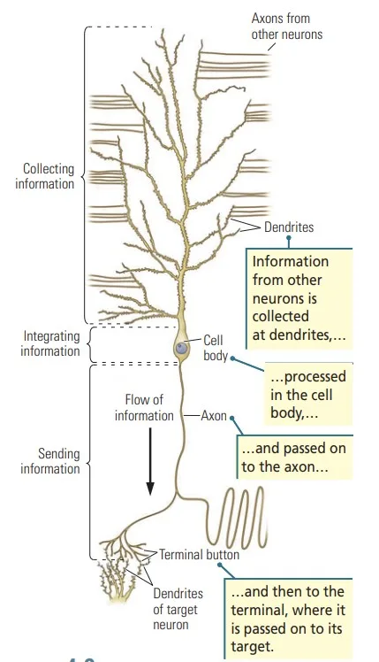
Flow of information through neuron
Processing and Regulation
While the flow of information appears linear- dendrites > soma > axon > terminal buttons – the neuron is not a simple conduit:
- It is an information-processing device.
- A neuron integrates thousands of inputs and produces a single output that reflects the summation of incoming signals.
- This ensures that the neuron communicates only the most relevant information to its target.
Watch : Structure function and types of neurons | Human Anatomy | 3D Biology
Conclusion
The structure of a neuron – comprising dendrites, the cell body (soma), the axon, and the terminal buttons is intricately designed to support its role in receiving, processing, and transmitting information. Each component contributes uniquely to neural communication, with dendrites gathering signals, the soma integrating inputs, and the axon transmitting outputs to other cells. Understanding the anatomy of a nerve cell is fundamental to comprehending how the brain and nervous system function. This knowledge not only provides insights into normal neural operations but also lays the groundwork for addressing neurological disorders, advancing therapies, and developing innovations in neuroscience and artificial intelligence.
Learn Types of neurons and Glial cells
Read more about Neuropsychology
References
- Kalat, J. W. (2019). Biological psychology. Cengage.
- Kolb, B., Whishaw, I. Q., & Teskey, G. C. (2016). An introduction to brain and behavior.
- Pinel, J. (2023). Biopsychology 10th Edition. Pearson.
Niwlikar, B. A. (2025, January 17). Structure of Neuron: Understanding Its anatomy and Role in Neural Communication. Careershodh. https://www.careershodh.com/structure-of-neuron/