
Introduction
The cerebral cortex of the human brain is divided into brain lobes that reflect both structural and functional specializations. These lobes—frontal, parietal, temporal, and occipital—coordinate complex processes such as motor control, sensory perception, language, memory, and executive function. Each lobe communicates with subcortical structures and other cortical areas, forming an intricate neural network essential for human behavior (Kalat, 2019; Pinel & Barnes, 2018).
Read More- Brain Hemispheres
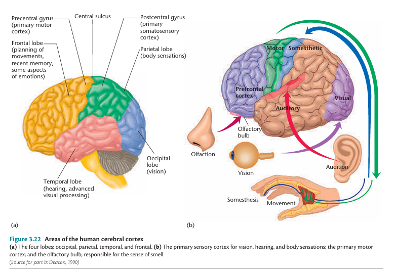
Brain Lobes
1. Frontal Lobe
The structure, function, and disorder of occipital lobe include-
Structure
The frontal lobe is the most anterior region of the cerebral cortex, bordered posteriorly by the central sulcus and inferiorly by the lateral sulcus. It encompasses the primary motor cortex (precentral gyrus), premotor cortex, prefrontal cortex, and, in the dominant hemisphere, Broca’s area.
Functions
Some functions associated include-
- Primary Motor Cortex (M1): Controls voluntary skeletal muscle movements via somatotopic organization (Pinel & Barnes, 2018).
- Prefrontal Cortex (PFC): Central to executive functions, including attention, working memory, problem-solving, inhibition, planning, and social behavior (Kalat, 2019).
- Broca’s Area: In the left hemisphere, this region is critical for speech production. Damage results in expressive aphasia (Pinel & Barnes, 2018).
- Orbitofrontal Cortex: Involved in decision-making, reward evaluation, and regulation of emotion and impulse control.
Disorders
Some disorders associated are-
- Frontal Lobe Syndrome: Characterized by apathy, disinhibition, poor judgment, and social inappropriateness due to PFC damage.
- Broca’s Aphasia: Results from lesions in Broca’s area; speech becomes non-fluent and effortful, though comprehension is preserved (Kalat, 2019).
- Attention Deficit Hyperactivity Disorder (ADHD): Often involves hypoactivity in the prefrontal cortex, impairing executive functioning (Pinel & Barnes, 2018).
- Schizophrenia: Frequently associated with reduced activity in the dorsolateral PFC, known as “hypofrontality” (Kalat, 2019).
2. Parietal Lobe
The structure, function, and disorder of occipital lobe include-
Structure
Located posterior to the frontal lobe and separated by the central sulcus, the parietal lobe includes the primary somatosensory cortex (postcentral gyrus) and regions involved in spatial awareness and integration of sensory input.
Functions
Some functions associated include-
- Somatosensory Processing: The postcentral gyrus receives tactile information from the body, including pain, temperature, and proprioception.
- Spatial Cognition: The right parietal lobe contributes to body awareness and visual-spatial integration (Pinel & Barnes, 2018).
- Numerical and Linguistic Processing: The left inferior parietal lobule assists in arithmetic and language functions (Kalat, 2019).
Disorders
Some disorders associated are-
- Hemispatial Neglect: Typically resulting from right parietal damage, this condition causes patients to ignore stimuli on the left side of their body or environment (Pinel & Barnes, 2018).
- Gerstmann’s Syndrome: Left parietal lesions may result in a constellation of deficits: agraphia, acalculia, finger agnosia, and left-right disorientation.

Gerstmann Syndrome
- Apraxia: Damage to the superior parietal lobule can lead to ideomotor or ideational apraxia, affecting coordinated movement planning.
3. Temporal Lobe
The structure, function, and disorder of occipital lobe include-
Structure
The temporal lobe lies beneath the lateral sulcus and includes structures such as the primary auditory cortex, Wernicke’s area, hippocampus, and amygdala.
Functions
Some functions associated include-
- Auditory Perception: The primary auditory cortex processes pitch, tone, and rhythm (Pinel & Barnes, 2018).
- Language Comprehension: Wernicke’s area, located in the left superior temporal gyrus, is crucial for interpreting spoken language.
- Memory and Learning: The hippocampus is vital for encoding and consolidating declarative memories (Kalat, 2019).
- Emotion: The amygdala evaluates emotionally salient stimuli and is important in fear conditioning.
Disorders
Some disorders associated are-
- Wernicke’s Aphasia: Fluent but nonsensical speech with impaired comprehension due to left temporal lobe damage (Pinel & Barnes, 2018).

Wernicke’s Aphasia
- Temporal Lobe Epilepsy: Causes focal seizures that may involve hallucinations, déjà vu, or automatisms.
- Anterograde Amnesia: Bilateral damage to the hippocampus, as in the case of H.M., prevents the formation of new episodic memories (Kalat, 2019).
- Prosopagnosia: Inability to recognize faces, typically from lesions in the right fusiform gyrus.
4. Occipital Lobe
The structure, function, and disorder of occipital lobe include-
Structure
The occipital lobe is the posterior-most part of the cortex, primarily devoted to processing visual stimuli. It includes the primary visual cortex (V1), also known as the striate cortex, and several visual association areas.
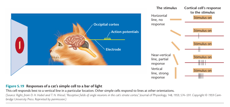
Hubel & Wisel’s Study on Visual Cortext
Functions
Some functions associated include-
- Basic Visual Processing: V1 processes information about light, edges, and motion from the retina.
- Ventral Stream (“What” Pathway): Projects to the temporal lobe and is involved in object and face recognition.
- Dorsal Stream (“Where” Pathway): Projects to the parietal lobe and manages spatial relationships and motion tracking (Pinel & Barnes, 2018).

What and Where Pathway
Disorders
Some disorders associated are-
- Cortical Blindness: Complete V1 damage can result in blindness despite intact ocular structures.
- Visual Agnosia: Impairments in recognizing objects due to ventral stream damage.
- Alexia Without Agraphia: Results from left occipital and corpus callosum damage; patients can write but cannot read.
Interconnectivity Between Lobes
The brain’s functionality depends not only on the individual lobes but also on their integration. For example:
- Reading a word requires occipital input (visual), parietal processing (spatial decoding), temporal interpretation (language), and frontal output (speech production).
- Conduction aphasia can occur from damage to the arcuate fasciculus, disrupting communication between Broca’s and Wernicke’s areas (Kalat, 2019).
Clinical Assessment and Imaging
Modern neuroimaging techniques, including fMRI, PET, and diffusion tensor imaging (DTI), provide real-time visualization of lobe-specific activity and white matter pathways. These tools are indispensable in localizing lesions in stroke, epilepsy, dementia, and traumatic brain injury (Pinel & Barnes, 2018).
Conclusion
The four cerebral lobes—frontal, parietal, temporal, and occipital—serve as functional domains within the brain, each contributing to the regulation of behavior, cognition, and sensory processing. Damage to specific lobes produces characteristic deficits that inform clinical diagnosis and therapeutic approaches. An integrative understanding of these lobes enhances our comprehension of the brain’s architecture and functionality, emphasizing the interplay of localized function and global brain dynamics.
References
Kalat, J. W. (2019). Biological psychology (13th ed.). Cengage Learning.
Pinel, J. P. J., & Barnes, S. J. (2018). Biopsychology (10th ed., Global ed.). Pearson Education.
Niwlikar, B. A. (2025, June 19). 4 Important Brain Lobes Their Structure, Functions, and Associated Disorders. Careershodh. https://www.careershodh.com/brain-lobes/